Fig. 5.
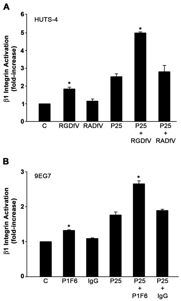
Activation of β1 integrin by β5 integrin blocking agents. A - MG-63 cells were pretreated for 45 minutes with 20 μM cyclic peptides RGDfV or RADfV and incubated with 50 μM of P25 or S25 in DMEM. Activation of β1 integrin was assessed by ELISA using the HUTS-4 antibody. B - MG-63 cells were preincubated for 2 hours with 20 μg/ml of normal mouse IgG, or β5 integrin blocking antibody P1F6 before treatment with P25 or S25. Integrin activation was assessed using monoclonal antibody 9EG7. All graphs show the levels of β1 integrin activation after normalization to total β1 levels as described in Fig. 1. Total β1 integrin did not change. Data represent one of three different experiments performed in triplicate. *Significantly different than control or P25-treated cells, respectively, t test, p < 0.05 (n=3).

