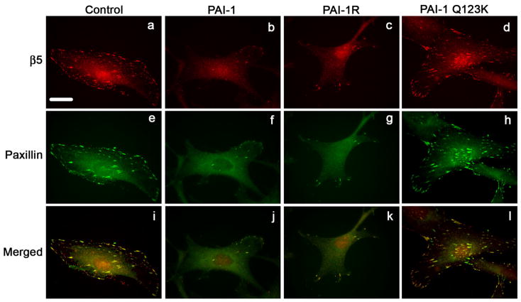
Fig. 6.
PAI-1 disrupts focal adhesions. MG-63 cell monolayers were plated overnight onto coverslips in complete media and then incubated for 3 hours with 20 nM of PAI-1 or PAI-1 mutants (PAI-1R and Q123K). Cells were subsequently fixed, permeabilized and stained for 1 hour with the clone 15F11 antibody for β5 integrin (panels a-d). After1 hour staining with the AlexaFluor594 derivatized anti-mouse secondary antibody, cells were washed and labeled for an additional hour with a paxillin-FITC antibody (panels e-h). Panels i-l show merged images indicating colocalization of β5 integrin with paxillin. Scale Bar, 20 μm.

