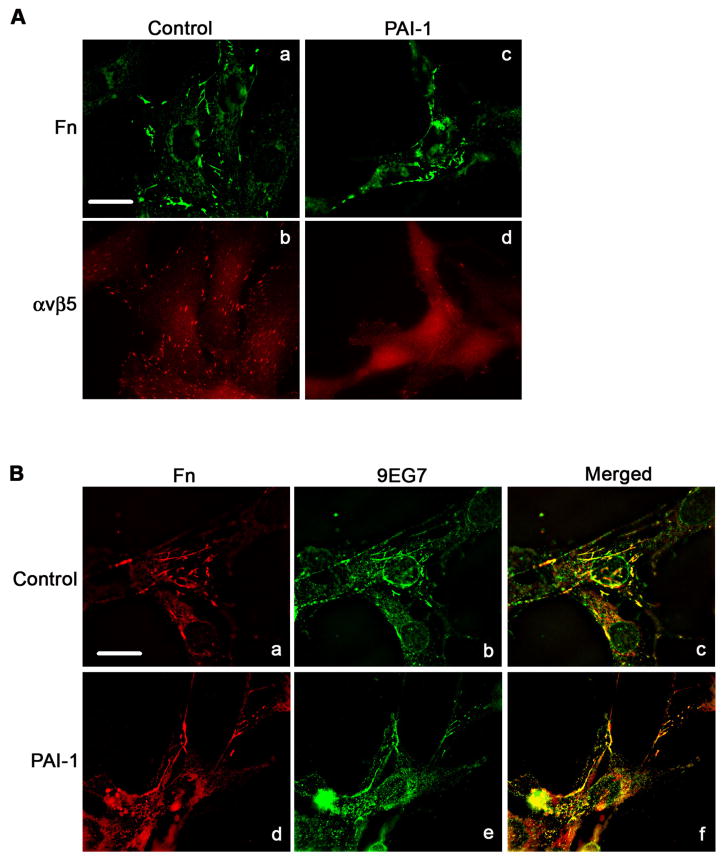
Fig. 9.
PAI-1 does not disrupt the association of α5β1 with fibronectin matrix. Cells were incubated for 3 hours in DMEM in the presence or absence of PAI-1, subsequently fixed, permeabilized and immunostained. A - Fibronectin matrix and β5 integrin were visualized by indirect immunofluorescence in non-treated cells (panels a and b) and PAI-1 treated cells (panels c and d). Scale Bar, 20 μm. B - Fibronectin and activated β1 integrin (detected with 9EG7 antibody) were visualized by indirect immunofluorescence in non-treated cells (panels a and b) and PAI-1 treated cells (panels d and e). Panels c and f shows merged pictures. Yellow staining indicates areas of colocalization of β1 integrin with fibronectin matrix. Scale Bar, 20 μm.

