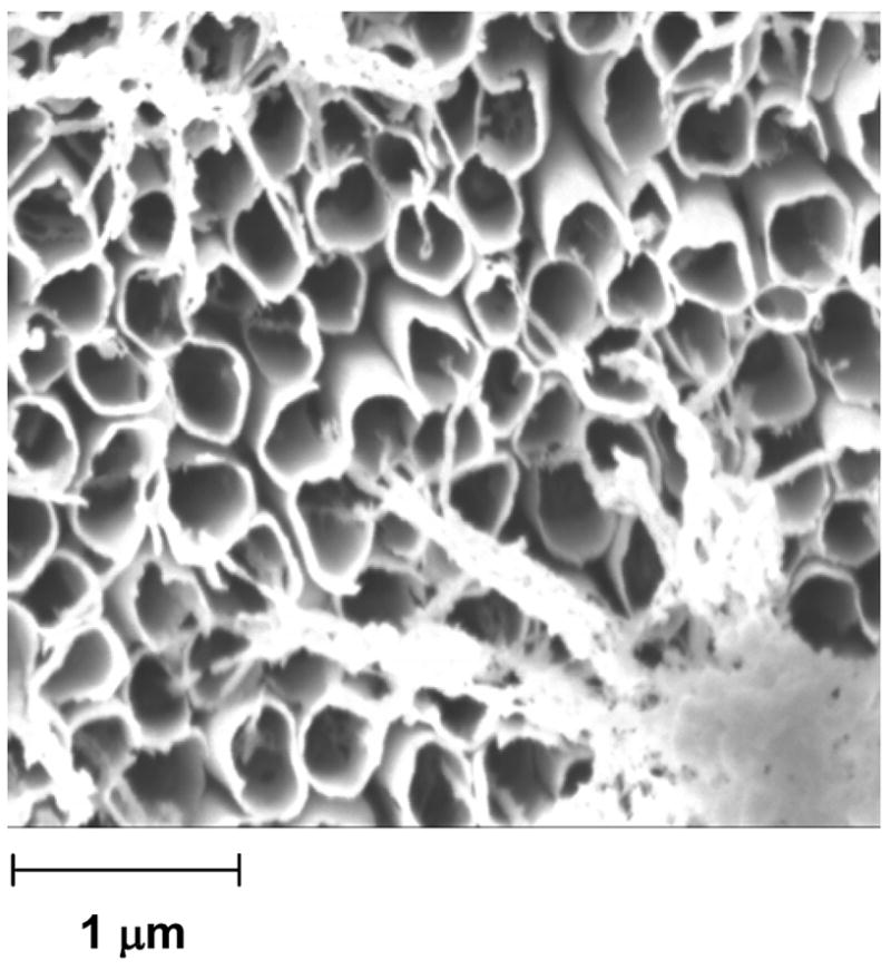
Figure 3.

SEM image of Au nanotubes (diameter = ~200 nm) produced when we treated the core-shell structures in Figure 1 with oxygen plasma for 5 min. A small amount of Au deposits inside of the PANI tube during step 2 of Scheme 1. We believe that the fibrous material in the bottom right and top left corners of the imageis Au that had deposited inside of the PANI tubes; this Au was released as the plasma etched the PANI, but remained tethered to the Au backing. Figure S1 shows a larger-area picture of the array of Au nanotubes.
