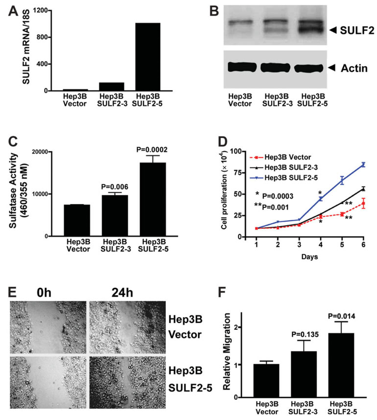
Fig. 2. Forced expression of SULF2 increases HCC cell proliferation and migration.
(A) Plasmids expressing the full-length SULF2 mRNA or the control empty vector pcDNA3.1 were stably transfected into Hep3B cells. SULF2 mRNA expression in different clones was confirmed with quantitative real-time RT-PCR in the three indicated clones. Compared to Hep3B Vector cells, Hep3B SULF2-3 cells showed an intermediate level of SULF2 mRNA expression, whereas Hep3B SULF2-5 cells showed a high level of SULF2 mRNA expression. (B) Western immunoblotting was performed on whole cell lysates from Hep3B Vector, Hep3B SULF2-3, and Hep3B SULF2-5 cells with antibodies against SULF2; the blots were then stripped and reprobed with antibodies against actin to control for loading. (C) Nonsteroid sulfatase activity was detected in the indicated clones after inhibition of steroid sulfatases with estrone-3-O-sulfamate. (D) One hundred thousand Hep3B Vector, Hep3B SULF2-3, or Hep3B SULF2-5 cells were plated into 6-well plates, and 3 wells of each cell clone were harvested, stained with trypan blue, and counted each day. The mean and standard error of triplicate measurements are plotted (P < 0.01 at days 4, 5, and 6, Hep3B SULF2-5 versus Hep3B Vector). (E) Scratch wounds were made in confluent cell culture monolayers with a 20-µL pipette tip; photomicrographs of the wounds were taken at 0 and 24 hours thereafter. (F) Relative migration of the wound edge was quantitated and showed a significantly increased rate of migration in Hep3B SULF2-5 cells when compared to Hep3B Vector cells (P = 0.013).

