
Figure 4.
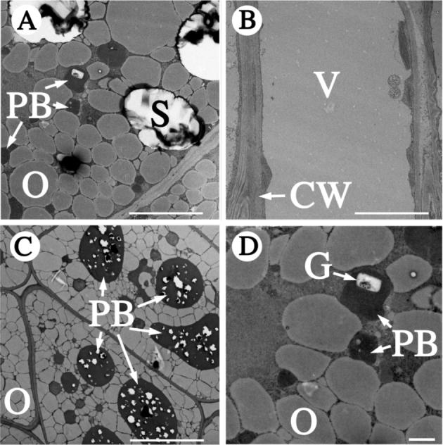
Pickle roots contain protein bodies and phytin globoids. The subcellular organization of a pickle root cell (panels A and D) was examined by transmission electron microscopy. Images from a wild-type root (B) and wild-type seed (C) are included for comparison. The larger protein bodies of seeds (C) contain numerous phytin globoids (white inclusions) that are not specifically marked as such. CW, cell wall; G, phytin globoid; O, oil body; PB, protein body; S, starch granule; V, vacuole. The white scale bars represent 10 μm in A, B and C or 1 μm in panel D.