
FIG. 2. Confocal images of a single mature skeletal muscle fiber.
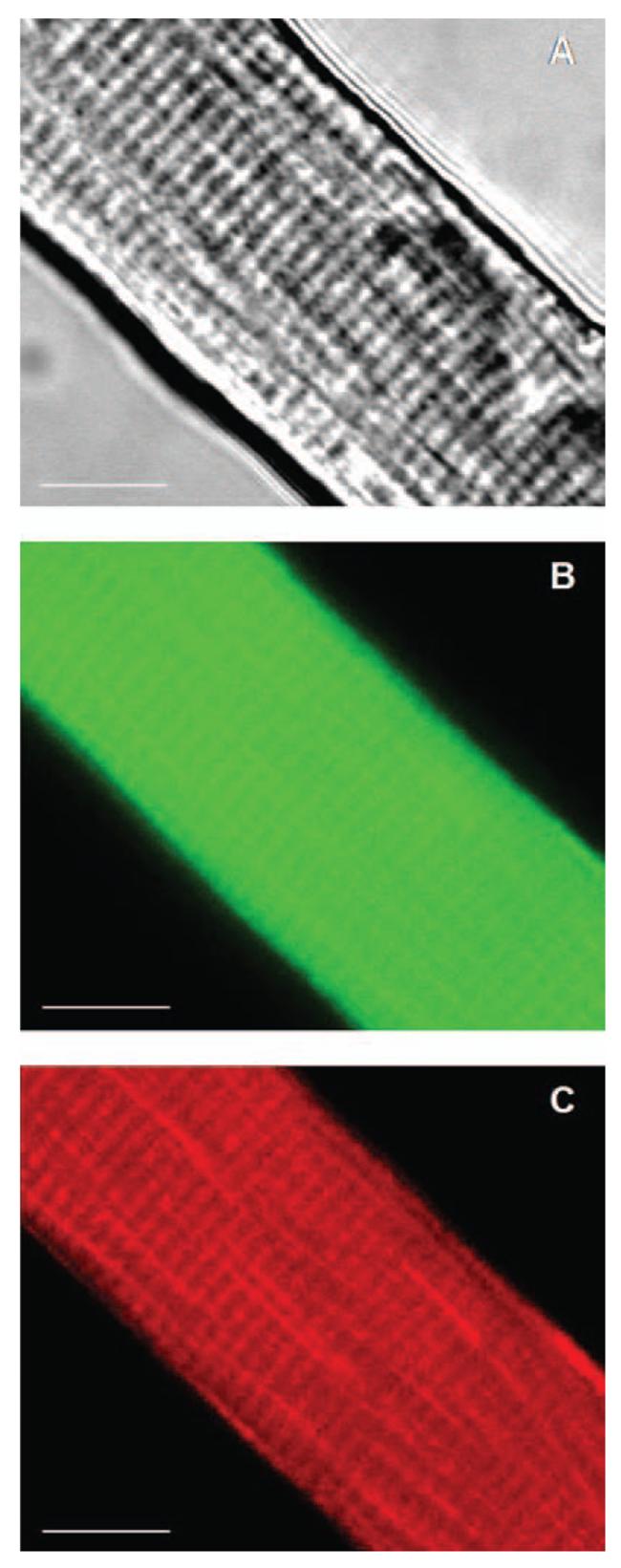
Selected area of a single mature skeletal muscle fiber loaded with the fluorophores CM-DCFH (17.5 μM) and Mitotracker red (20 nM). (A) Bright-field image. (B) and (C) Confocal images of the same optical section (thick = 2.61 μm) showing: CM-DCF fluorescence (B) and Mitotracker red fluorescence (C). 63× original magnification. Scale bar 10 = μm.