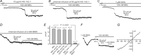
Figure 3. Melatonin increases [cGMP]i most likely via inhibiting PDE.
A, melatonin of 100 nm induced a current, which was comparable in density to that recorded in Ringer solution when the cell was incubated with 10 μg ml−1 HS-142-1 for 10 min. B, during internal infusion of 50 μg ml−1 HS-142-1, melatonin persisted to induce a current of comparable density. C, following preincubation of 1 μm ODQ for 10 min, melatonin still induced an inward current with comparable density. D, internal infusion of 2 mm IBMX induced an inward current. When the current became steady, addition of 100 nm melatonin induced a current of much less density. E, bar chart, showing that neither HS-142-1 nor ODQ changed the melatonin currents, but IBMX did. P = 0.518 and 0.488 for perfusion and internal infusion of HS-142-1, respectively; P = 0.461 for ODQ; P < 0.0001 for IBMX. F, puff of 1 mm IBMX to the dendrites of a Rod-ON-BC for 15 s induced an inward current. When incubated with 100 nm melatonin for about 1.5 min, the current induced by IBMX was much smaller in density. G, I–V curve of the IBMX current induced from a Rod-ON-BC, using a voltage ramp from −80 mV to 40 mV for 500 ms. The curve yields an Erev of –4.2 mV.

