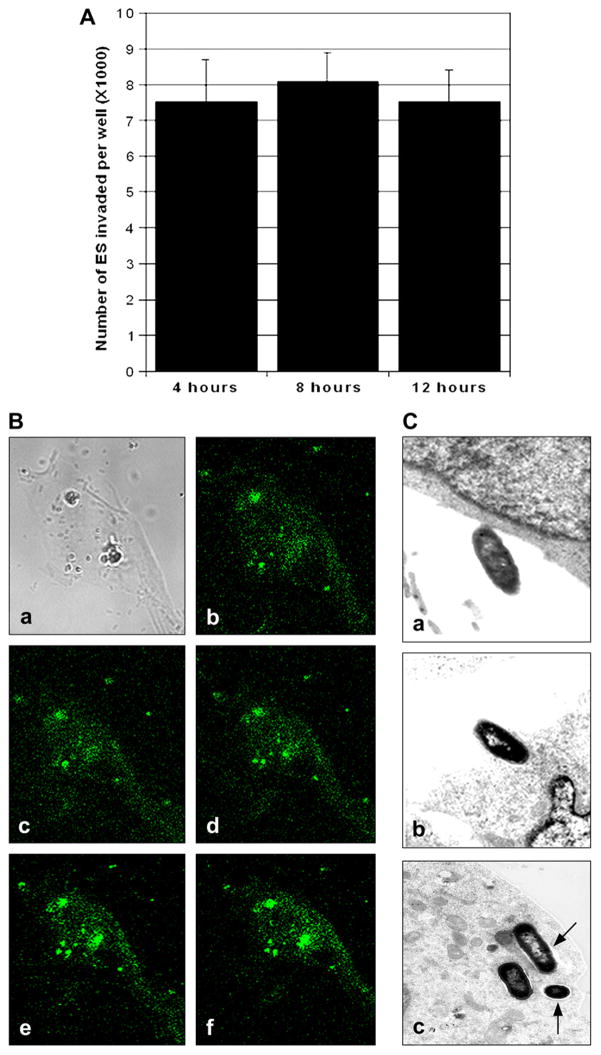
Fig. 2.
Internalized ES does not multiply inside HBMEC. (A) HBMEC were infected with ES for 4 h followed incubation with gentamicin containing medium for additional 4 h and 8 h. The number of intracellular bacteria was determined as described in Fig. 1. The data represent means ± SD from three independent experiments performed in triplicate. (B) HBMEC monolayers were infected with GFP+ ES for 4 h, washed, and fixed with 2% paraformaldehyde. z-Stacks of confocal images were acquired using a Leica confocal laser-scanning microscope and Leica confocal software equipped with 63× oil lens. (C) In separate experiments, HBMEC monolayers were infected with 106 cfu/well for varying periods. Samples were taken at 1 h (a), 2 h (b), and 8 h (c) post-infection and were processed for transmission electron microscopy as described in Section 4. Arrows indicate the vacuoles containing ES. Magnification for the included photographs: (a, b) 12,000; (c) 13,000 and (d) 8500.

