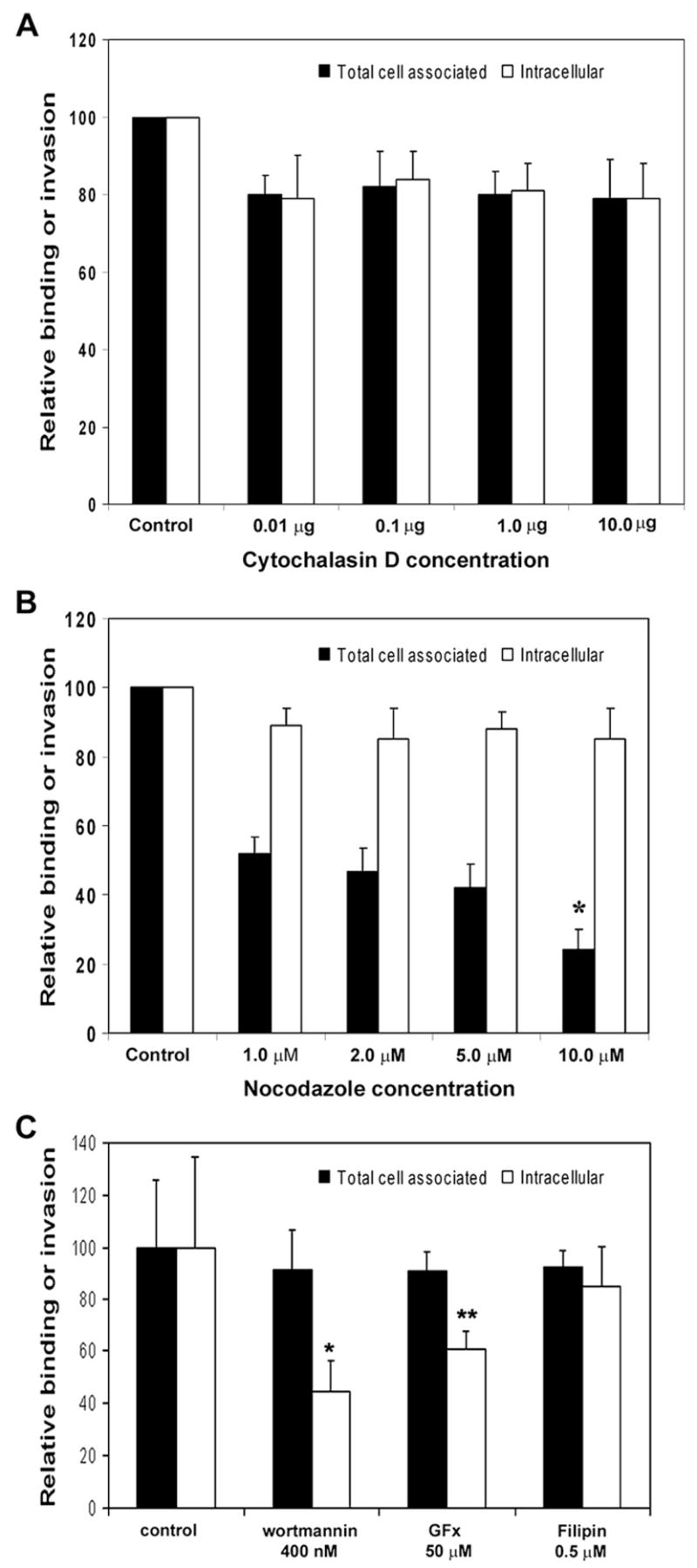
Fig. 4.

Effect of various inhibitors on ES invasion of HBMEC. Different concentrations of cytochalasin D (A), nocodazole (B) or wortmannin, GFx and filipin (C) were added to confluent monolayers of HBMEC for 30 min at 37 °C prior to the addition of ES. Total cell associated and intracellular ES was determined as described earlier. The experiments were performed at least 3 times in triplicate and are expressed as means ± SD. *p < 0.001 and **p < 0.05 by Student’s t test.
