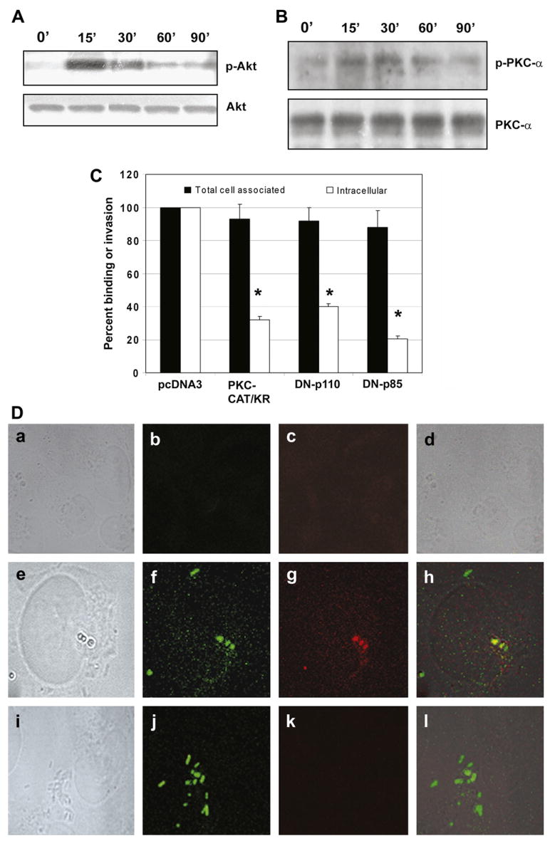
Fig. 6.
ES incubation of HBMEC activates PI3-kinase and PKC-α. Confluent monolayers of HBMEC were infected with ES (106 cfu/well) for 0–90 min, total cell lysates prepared, and subjected to Western blotting with anti-phospho-Akt or anti-Akt antibodies (A). Similar blot was also subjected to Western blotting with anti-phospho-PKC-α or anti-PKC-α antibody (B). In addition, HBMEC were transfected with dominant negative mutants of either p85 subunit of PI3-kinase or PKC-α. Total cell associated (binding) and intracellular bacteria (invasion) were determined relative to the binding and invasion of HBMEC being taken as 100%. The experiments were performed at least 3 times in triplicate and the data are expressed as means ± SD (C). The invasion of ES into HBMEC transfectants was significantly lower than into HBMEC transfected with pcDNA3, *p < 0.001 by Student’s t test. (D) HBMEC, non-transfected (a–d), transfected with plasmid alone (e–h) or with DN-PKC-α were either non-infected (a–d) or infected with GFP+ ES for 90 min (e–l). The cells were washed, fixed, and stained with anti-phospho-PKC-α antibody (c, g, and k). The bright light images were taken to show the morphology and boundaries of the cells (a, e, and i). The overlay images showed that ES entry induced the accumulation of phospho-PKC-α (yellow color) in HBMEC.

