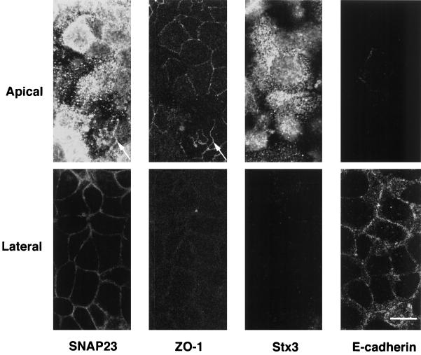
Figure 4.
Localization of t-SNARES in horizontal confocal sections of CaCo-2 cells. Double immunofluorescence micrographs of CaCo-2 cells stained for SNAP23 and ZO-1, a tight junction marker (left) or syntaxin 3(Stx3) and E-cadherin, a basolateral marker (right). Syntaxin 3 is restricted to the apical plasma membrane (note the lack of staining of the lateral plasma membrane that is positive for E-cadherin in bottom right micrographs). SNAP23 is highly concentrated at the apical plasmalemma and is present in tight junctions (top left micrographs; arrow indicates colocalization with ZO-1) and at low level in the lateral plasma membrane below tight junctions (bottom left). Bar, 10 μm.

