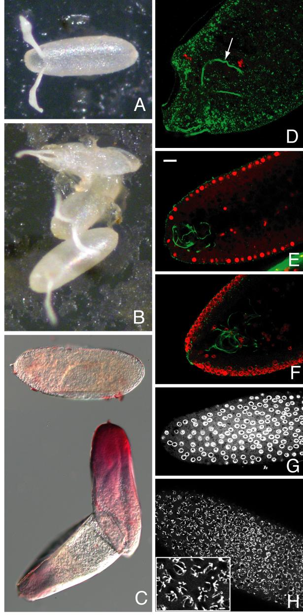
Fig. 4. Vitelline membrane integrity and embryonic development are compromised by psd knockdown.
(A) OR and (B) psd knockdown laid eggs showing occasional collapse of the latter (top egg in B). (C) Most psd knockdown eggs are permeable to the dye neutral red following bleach dechorionation; top dye-impermeable egg was imaged in a different field of the same slide. (D-H) Embryos stained with propidium iodide to label DNA (red in D-F, white in G and H), and with DROP1.1 antibody to label the sperm tail (green in D-F only). (D) Severely affected psd knockdown embryo showing only oocyte and sperm DNA is still fertilized (sperm tail marked with arrow). (E) Sagittal section of wild-type syncytial blastoderm stage embryo showing even propidium iodide staining throughout nuclei. (F) Sagittal section of syncytial blastoderm stage psd knockdown embryo showing ring-like propidium iodide staining at periphery of nuclei. (G-H) Surface views of syncytial blastoderm stage psd knockdown embryos fixed after 0-4 hour collections to minimize effects of degradation after developmental arrest. These show either clear ring-like staining of DNA (G) or visibly separate chromosomes that are also at the nuclear periphery (H, enlarged in inset). Bar equals 20 μm in E-H, 10 μm in D.

