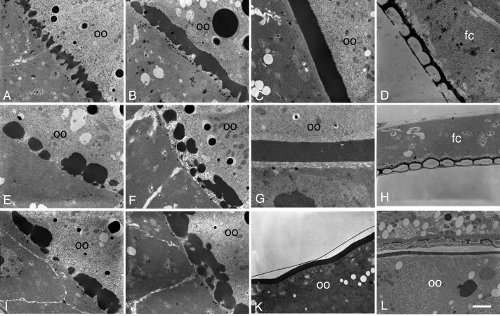
Fig. 5. Electron microscopy reveals early defects in vitelline membrane morphogenesis in the psd knockdown.
(A-D) OR egg chambers. (A) Stage 10A, uniform array of vitelline bodies in the perivitelline space between the oocyte (oo) and follicle cells. (B) Stage 10B, coalescence of vitelline bodies. (C) Stage 11, uniform vitelline membrane. (D) Late-stage egg chamber, showing the endochorion, consisting of a thin floor and thick roof separated by pillars and spaces containing only light flocculent material. Follicle cells are indicated (fc), and the chorion has separated from the oocyte and vitelline membrane, which are out of the field of view. (E-K) psd knockdown egg chambers. (E, I) Stage 10A, irregularly sized and distributed vitelline bodies. and abnormally large gaps between them. (F, J) Late Stage 10A or Stage 10B, coalescence of vitelline bodies. (G) Stage 11, continuous vitelline membrane that can be thinner than in wild type, and that has small holes but no full-thickness gaps. (H, K) Late-stage egg chambers show a mature vitelline membrane with normal thickness but persistent holes (K), and normal inner chorionic layer (thin line above vitelline membrane in K) and endochorion (H). In a sV23-null mature egg chamber (L), an initially normal appearing endochorion has collapsed, with dense flocculent material in the endochorionic spaces and with electron-dense material scattered throughout a thin vitelline membrane (Pascucci et al., 1996). Bar equals 2 μm in all images except D, 750 nm, and G, 1 μm.

