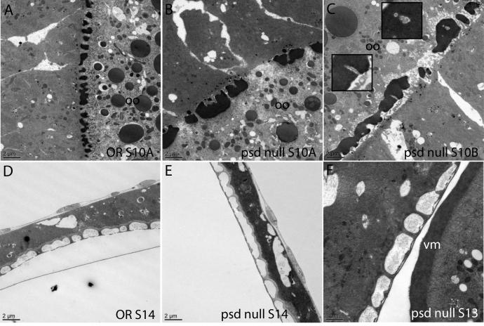
Fig. 6. Electron microscopy of the psd null confirms disorganized vitelline membrane assembly and intact chorion architecture.
Comparison of OR (A) and psd null (B) Stage 10A egg chambers illustrates the “boulders and gaps” appearance of the vitelline bodies in the psd null. During coalescence of the vitelline membrane in Stage 10B, there appeared to be trapping of microvilli within the vitelline membrane in the psd null (C; insets show enlarged images). The psd null egg chambers showed normal architecture of the chorion layers (E, F) as compared to OR (D), and the final vitelline membrane structure did not contain gaps. Bars equal 2 μm in all images except F, 1 μm.

