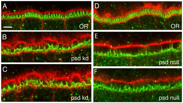
Fig. 7. Loss of Palisade results in disruption of follicle cell microvillar organization during vitelline membrane synthesis.
Shown are close-up views of the perivitelline space from six different stage 10A egg chambers from OR (A, D), psd knockdown (B, C), and psd null (E, F) females. The cortical actin cytoskeleton of the oocyte (top portion of each panel) and follicle cell epithelium (bottom portion) is stained with Texas Red-phalloidin (red), while the follicle cell microvilli are stained with a Cad99C antibody (green). Bar equals 5 μm.

