Figure 5.
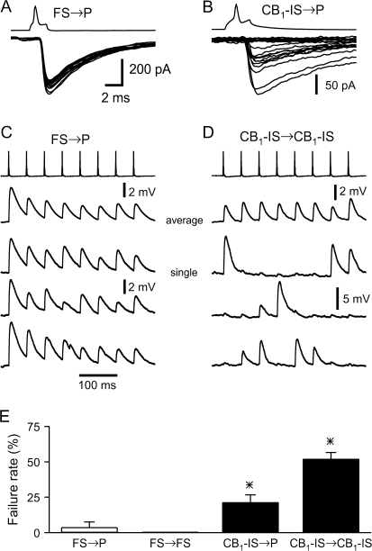
Synaptic responses mediated by CB1-IS cells show much more variability than those mediated by FS cells. (A) Superimposition of 15 consecutive IPSCs in a pair between a presynaptic FS cell and a postsynaptic pyramid ([Cl−] = 130 mM) in layer 2/3. Note the lack of failures. (B) Superimposition of 20 consecutive IPSCs in a pair between a presynaptic CB1-IS cell and a postsynaptic pyramid ([Cl−] = 130 mM) in layer 2/3. Note that about half of the responses are failures. (C) Connection between a presynaptic FS cells and postsynaptic pyramid in layer 5. Both cells are under current clamp. Trains of 8 action potentials at 20 Hz were generated in the FS cell every 5 s. Depolarizing IPSPs were recorded from the postsynaptic cell ([Cl−] = 40 mM, Vm = -90 mV). The top trace shows the average of 20 trials. The 3 lower traces are individual trials. (D) Connection between 2 CB1-IS cells in layer 2/3 that was also electrically coupled. Both cells are under current clamp. Trains of 8 action potentials at 20 Hz were generated in the presynaptic CB1-IS cells every 6 s. Depolarizing IPSPs were recorded from the postsynaptic cell ([Cl−] = 130 mM) at resting potential. The top trace shows the average of 50 trials. The 3 lower traces are individual trials. Note that many presynaptic action potentials failed to produce a postsynaptic potential. However, the weak electrical coupling produced small postsynaptic depolarization following each presynaptic spike. (E) Summary data of failure rate from the 4 types of connections (FS → P, 3.8% n = 5 pairs; FS → FS, 0% n = 6 pairs; CB1-IS → CB1-IS, 52% n = 12 pairs and CB1-IS → P, 21% n = 9).

