Figure 2.

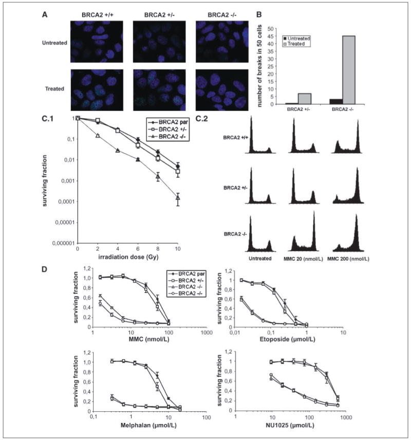
Characterization of BRCA2-null cells. A, immunofluorescence of RAD51 nuclear foci in untreated and treated cells (2.4 μg/mL MMC). BRCA2 +/−, BRCA2wt/Δex11; BRCA2−/−, BRCA2Δex11/Δex11. B, analysis of 50 metaphases for chromosomal aberrations in untreated and treated (equitoxic doses of MMC) heterozygous and knockout clones. C.1, colony formation assays on irradiation. BRCA2 par, parental BRCA2wt/mut × 2. Points, mean of three independent experiments; bars, SE. C.2, cell cycle analysis 48 h after MMC treatment. Representative cell cycle profiles obtained after treatment with indicated concentrations of MMC. D, cell proliferation following treatment with selected drugs. Two BRCA2 −/− (BRCA2Δex11/Δex11) subclones were analyzed. Points, mean of three independent experiments; bars, SE.
