Figure 3.
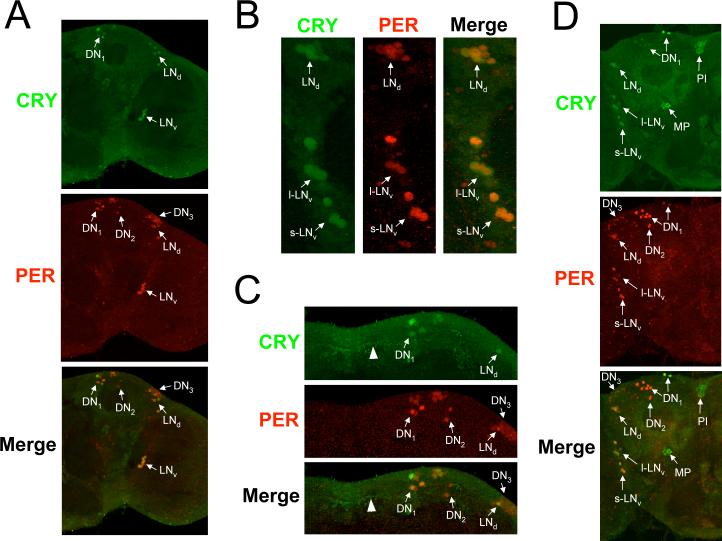
CRY expression in adult brains. Brains were dissected from w flies collected at CT19 on the third day of DD, immunostained with CRY (GP23) and PER antisera, and imaged via confocal microscopy. A minimum of six brains were analyzed with similar results. A. Images of a 2μm z-stack projection through the left hemisphere are shown, where dorsal is at the top. Colocalization of CRY (green) and PER (red) immunofluorescence is seen in the merged image as yellow. Arrows indicate CRY and/or PER immunostaining in oscillator neurons. s-LNv, small ventrolateral neurons; l-LNvs, large ventrolateral neurons; LNds, dorsolateral neurons; DN1, dorsal neuron1s; DN2, dorsal neuron 2s; DN3, dorsal neuron 3s. B. A compressed stack of six 1 μm optical sections through the lateral protocerebrum is shown. Colocalization of CRY (green) and PER (red) immunofluorescence is seen in the merged image as yellow. Arrows indicate CRY and/or PER immunostaining in oscillator neurons. Oscillator neurons are labeled as in panel A. C. A compressed stack of six 1μm optical sections through the dorsal protocerebrum is shown. Colocalization of CRY (green) and PER (red) immunofluorescence is seen in the merged image as yellow. Arrows indicate CRY and/or PER immunostaining in oscillator neurons. Oscillator neurons are labeled as in panel A. D. Images of a 2μm z-stack projection through the right hemisphere are shown, where dorsal is at the top. CRY and PER immunostaining and oscillator cells are as described in panels A-C, CRY-IR is also detected in the Pars Intercerebralus (PI) and the medial protocerebrum (MP).

