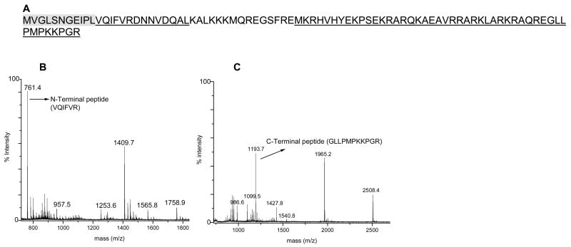
Figure 9.
(A) Sequence coverage map of N-terminally truncated S21. Underlined residues indicate the peptide masses observed. The shaded sequence is the absent N-terminal undecapeptide (see text for discussion). (B) MALDI spectrum of ribosomal protein S21 tryptic digest. (C) MALDI spectrum of ribosomal protein S21 Glu-C digest.

