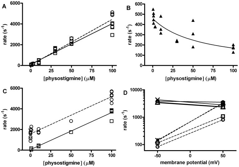
Figure 8. Properties of channel block by physostigmine.
The increase in the concentration of physostigmine was found to result in reduced open duration and an increase in the rate of entry into a ∼3-7 ms closed state. Both observations are consistent with physostigmine-induced channel block. (A) The inverse of the open duration (circles and dashed line) and the rate of entry into the putative blocked state (squares and solid line) for εT264P mutant receptors are plotted as a function of physostigmine concentration. The lines were fitted to rate = rate at no physostigmine + k+B* · [physostigmine], where k+B* is the apparent blocking rate. The estimates for k+B* were 44 ± 2 μM-1s-1 when fitting the reduction in the open duration, and 39 ± 2 μM-1s-1 when fitting the increase in the rate of entry into the putative blocked state. (B) The relationship between the inverse of the duration of the putative blocked state and physostigmine concentration in the εT264P mutant receptor. The increase in the duration of the putative blocked state at higher physostigmine concentrations is consistent with the presence of two (or more) blocking sites per receptor. The line was fitted to 1/τBlocked = k-B (2k-B / (2k-B + k+B* · [physostigmine])). This equation assumes the presence of two, equivalent blocking sites per receptor. The fitting results are: k+B = 15.6 ± 4.8 μM-1s-1, and k-B = 458 ± 28 s-1. (C) The inverse of the open duration (circles) and the rate of entry into the putative blocked state (squares) for the wild type receptor activated by 1 mM carbachol in the presence of physostigmine are plotted as a function of physostigmine concentration. The estimates for k+B* were 36 ± 2 μM-1s-1 when fitting the reduction in the open duration, and also 36 ± 2 μM-1s-1 when fitting the increase in the rate of entry into the putative blocked state. (D) Changes in membrane potential strongly affected the rate of return from the blocked state but were ineffective at altering the rate of entry into the blocked state. The values for k+B* (solid lines) and k-B* (dashed lines) in the presence of 100 μM physostigmine were estimated for wild type receptors activated by 1 mM carbachol (circles) or 200 μM ACh (squares), εT264P receptors activated by 100 μM carbachol (triangles), and εT264P receptors exposed solely to physostigmine (crosses) at -50 mV and +50 mV membrane potential. For the rate of development of block, the H value (change in membrane potential needed for an e-fold change in parameter) was 1141 mV (wild type + carbachol), 268 mV (wild type + ACh), 261 mV (εT264P + carbachol), or 227 mV (εT264P + physostigmine alone). For the rate of recovery from block, the H value was 44 mV (wild type + carbachol), 45 mV (wild type + ACh), 31 mV (εT264P + carbachol), or 32 mV (εT264P + physostigmine alone). For panels A through C, each point shows data from one patch, while for D each point shows results from the combined analysis of data from 2 or 3 patches.

