Fig. 5.
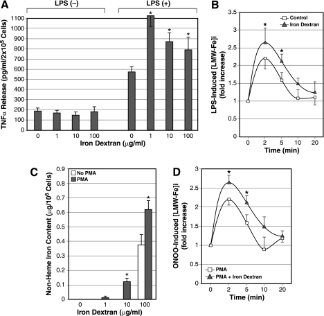
A: normal rat cultured KC were pretreated with the different concentrations of iron dextran and stimulated with LPS to determine the effects of iron loading on TNF-α release. Note iron dextran treatment alone does not affect the basal TNF-α release (left) but enhances LPS-stimulated TNF-α release twofold at 1 μg/ml concentration and 40–50% at 10–100 μg/ml of iron dextran (right). *P < 0.05 compared with the cells without the iron dextran treatment. B: iron loading by incubation with 1 μg/ml iron dextran overnight accentuates the ILI response in cultured KC as determined by LPS-induced [LMW-Fe]i. *P < 0.05 compared with the cells without prior iron dextran treatment (control). C: PMA-induced macrophages from rat blood monocytes take up iron dextran to increase iron storage more efficiently than vehicle-treated monocytes (no PMA). Normal rat blood monocytes were cultured with or without PMA (100 nM) and subsequently treated with iron dextran for 24 h before the cells were collected for determination of nonheme iron content. *P < 0.05 compared with vehicle-treated rat blood monocytes (no PMA). D: iron dextran loading enhances peroxynitrite-induced ILI response in rat blood monocyte-derived macrophages by PMA treatment. PMA-induced rat macrophages with or without iron dextran treatment (1 μg/ml) were analyzed for peroxynitrite-induced [LMW-Fe]i using Fe59Cl. *P < 0.05 compared with the cells without iron dextran treatment.

