Fig. 2.
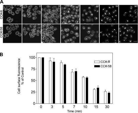
Agonist-induced CCK receptor internalization. A: representative confocal microscopic images demonstrating internalization of yellow fluorescent protein (YFP)-tagged CCK1 receptors expressed on Chinese hamster ovary (CHO) cells after their occupation with nonfluorescent CCK peptides. Time points after stimulation are noted. Like CCK-8, CCK-58 stimulated prompt and extensive CCK receptor internalization, with similar patterns of distribution. The kinetics of internalization were also similar after stimulation with the 2 CCK peptides. Scale bar = 25 μm. B: quantitation of the receptor on the cell surface under these conditions. Results reflect means ± SE of data from 3 independent experiments.

