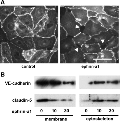
Fig. 5.
A: immunofluorescence microscopy of hLMVEC shows loss of claudin-5 staining at cell-cell junctions and endothelial gap formation (arrowheads) after 30 min of ephrin-a1 (2.5 μg/ml) stimulation. Scale bar, 15 μm. B: ephrin-a1 stimulation of hLMVEC (0, 10, or 30 min) causes subcellular redistribution of claudin-5 and VE-cadherin out of membrane fraction and into cytoskeletal fraction.

