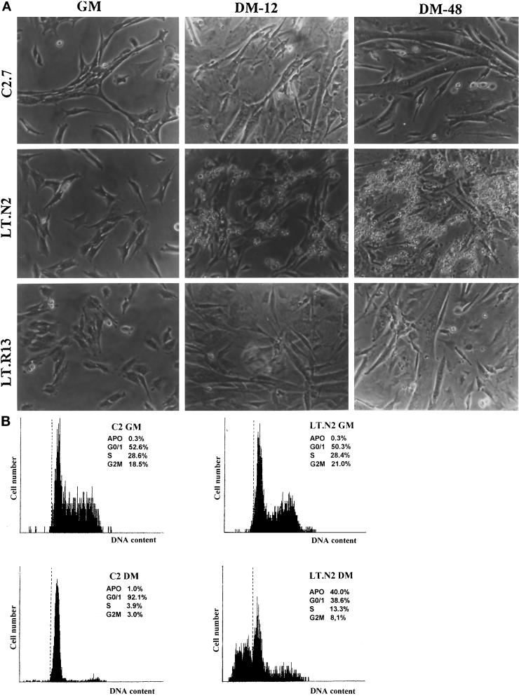
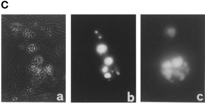
Figure 1.
PyLT-expressing myoblast undergo apoptosis after growth factor withdrawal. (A) Phase contrast micrographs showing the appearance of C2.7, LT.N2, and LT.R13 cells during exponential growth (GM) or 12 and 48 h after the shift to differentiation medium (DM-12 and DM-48, respectively). (B) Flow cytometric analysis of acridine-orange-stained LT.N2 cells compared with the parental C2.7 cells (C2), showing their cell cycle profile in growth medium (GM) and 24 h after the shift to differentiation medium (DM). (C) (facing page) Phase contrast (a) and fluorescence micrographs (b and c) of ethidium-bromide–stained LT.N2 cells, showing the nuclear morphology of dying cells (panel c represents a magnification of an apoptotic nucleus).


