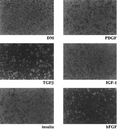
Figure 2.
Apoptosis in PyLT-expressing myoblasts cells is inhibited by bFGF and TGFβ. Phase contrast micrographs of LT.N2 cells 24 h after the shift to differentiation medium, either alone or supplemented with each of the indicated growth factors: PDGF (50 ng/ml), TGFβ (10 ng/ml), IGFI (50 ng/ml), insulin (10 μg/ml), or bFGF (50 ng/ml).

