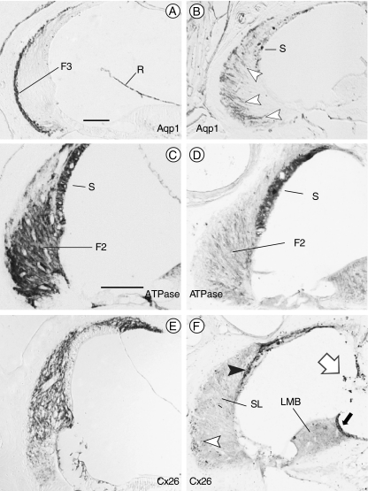
FIG. 3.
Spiral ligament fibrocytes are abnormal in mutant mice. Immunostaining of wild type (A, C, and E) and Brn4−;Tbx1+/− (B, D, and F) inner ears. A Normal distribution of type III fibrocytes (F3) at the junction of the spiral ligament and the petrous bone immunostained for Aquaporin 1 (Aqp1). B Aqp1 immunostained cells in Brn4−;Tbx1+/− ears are situated broadly throughout the spiral ligament (arrowheads). C Type II fibrocytes (F2) in the spiral ligament express Na+, K+–ATPase in wild type inner ears. The stria vascularis (S) also exhibits positive immunostaining. D The type II fibrocytes in Brn4−;Tbx1+/− mice only weakly express Na+, K+–ATPase; however, the stria remains strongly positive. E Connexin 26 (Cx26) is expressed broadly in the spiral ligament of a wild type inner ear. F Cx26 immunostaining of the inner ear of a Brn4−;Tbx1+/− mouse shows only a few positive cells within the spiral ligament (open arrowhead), but strongly stains the strial basal and intermediate cells (closed arrowhead). The spiral limbus (LMB) expresses Cx26 in wild type mice (not shown); however, it fails to stain in Brn4−;Tbx1+/− mice save for a dark strip of supralimbal cells (closed arrow). Melanocytes stain nonspecifically (open arrow). F2, type II fibrocytes; F3, type III fibrocytes; R, Reissner’s membrane; S, stria vascularis; SL, spiral ligament; and LMB, spiral limbus. Scale bar is 100 μm.

