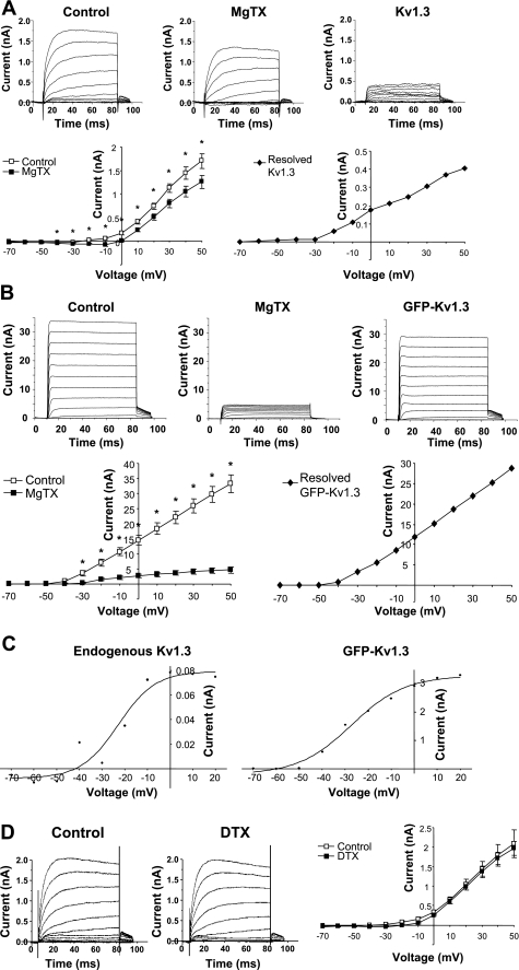
Fig. 4.
Voltage-clamp analyses of Kv1.3 in postganglionic sympathetic neurons. A: ionic current measured in untransfected dissociated sympathetic neurons in the absence (control; n = 18) and presence of 1 nM margatoxin (MgTX; n = 15). Current traces for each condition represent the average of unpaired measurements made in multiple cells. *Significant difference between MgTX and control (P ≤ 0.05; unpaired t-test). Resolved Kv1.3 current was obtained by subtracting MgTX from control. B: ionic current measured in dissociated sympathetic neurons transfected with GFP-Kv1.3 in the absence (control; n = 3) and presence of 1 nM MgTX (n = 3). Current traces for each condition represent the average of unpaired measurements made in multiple cells. *Significant difference between MgTX and control (P ≤ 0.02; unpaired t-test). Resolved GFP-Kv1.3 current was obtained by subtracting MgTX from control. C: activation curves were plotted from the tail current of endogenous Kv1.3 and GFP-Kv1.3 and fit to a Boltzmann function. D: ionic current measured in dissociated sympathetic neurons in the absence (control; n = 10) and presence of 100 nM DTX (n = 18; P > 0.05).

