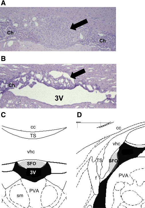
Fig. 1.
Histological sections and schematic diagrams at the level of the subfornical organ (SFO). Representative coronal sections at the level of the SFO (arrows) from a sham-lesioned (A) and an SFO-lesioned rat (B) and corresponding coronal (C) and sagittal diagrams (D) are shown. All rats included in the final data analysis had lesions encompassing the stippled areas shown on the schematic. Staining in A and B is hematoxylin and eosin (×40 magnification). 3V, third ventricle; cc, corpus callosum; f, fornix; PVA, anterior part of the paraventricular thalamic nucleus; sm, stria medullaris; TS, triangular septal nucleus; vhc, ventral hippocampal commissure; Ch, choroid plexus. Diagrams in C and D are modified from the rat brain atlas of Paxinos and Watson (38a).

