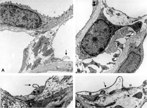
Fig. 9.
Ultrastructural patterns of vasa recta areas, inner stripe. A: control picture showing a thin limb on top and 2 capillary sections from vasa recta area. The capillary on the right shows a single detached endothelial blister. B–D: high-dose l-NMMA. B: centrally positioned fibroblast with dilated rough endoplasmic reticulum. Neighboring thin limb epithelia reveal layered basement membranes. C and D: vasa recta walls showing capillary detachments of fenestrated endothelium of different degrees.

