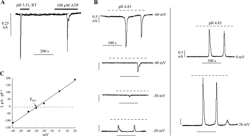
Fig. 2.
pH-evoked current in suburothelial myofibroblasts. A: membrane current in response to a low-pH solution and 100 μM ATP (Vh −60 mV). B: membrane current in response to exposure to a superfusate of pH 4.83. In the different panels, the Vh of the cell was held at the various values indicated. C: plot of the peak current as a function of Vh for the data shown in B. Erev, reversal potential.

