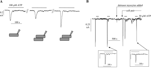
Fig. 4.
A: membrane currents elicited by 100 μM ATP in an isolated cell (left), when touching loosely on a second cell (middle), and more firmly on the second cell (right). Bottom: drawing of the experimental system. B: continuous tracing of membrane current showing inward transients elicited by repeated exposures to 30 μM ATP. At the first arrow, isolated detrusor myocytes were also added to the cell chamber; at the second arrow, the myofibroblast from which recordings were made was pressed firmly against a smooth muscle cell. Vh, −60 mV. Insets: second and fourth ATP-evoked current transient on a faster time base.

