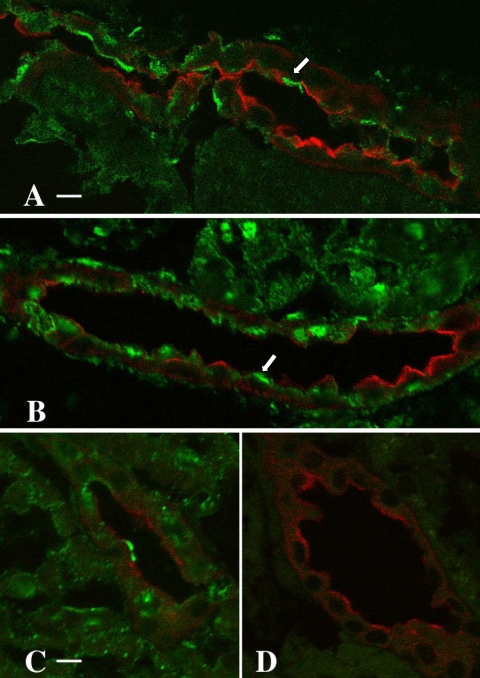
Fig. 3.
Immunolocalization of BK α-subunit in the kidney of adult rabbits maintained on LS (A, C, D) and HS (B) diets. Cryosections of kidneys from LS- and HS-fed animals were colabeled with CY3-conjugated Dolichos biflorus agglutinin (DBA; red), a principal cell marker, and an anti-BK channel α-subunit antibody, visualized with an FITC-labeled (green) secondary antibody. The pattern of apical expression, localized almost exclusively to DBA-negative cells (arrows), was similar in both experimental groups (compare A and B, same magnification; scale bar in A = 10 μm). Two sections of LS kidney cortex (C and D, same magnification) were labeled on the same day, one with the anti-BK channel antibody (C; scale bar = 5 μm) and the other (D) with antibody in the presence of excess immunizing peptide. Preincubation of the BK channel antibody with the immunizing peptide completely abolished anti-BK channel antibody labeling (D).

