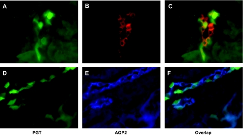
Fig. 5.
GFP-positive cells are PGT-expressing cells. Fluorescence microscopy of kidney cryosections of a PGT-GFP transgenic mouse (A and D), immunofluorescence labeling for PGT using Alexa Fluor 568 (red, B and E). C and F are the merges of A and B, and D and E, respectively. A, B, and C are by 10× magnification; D, E, and F are 20× magnification.

