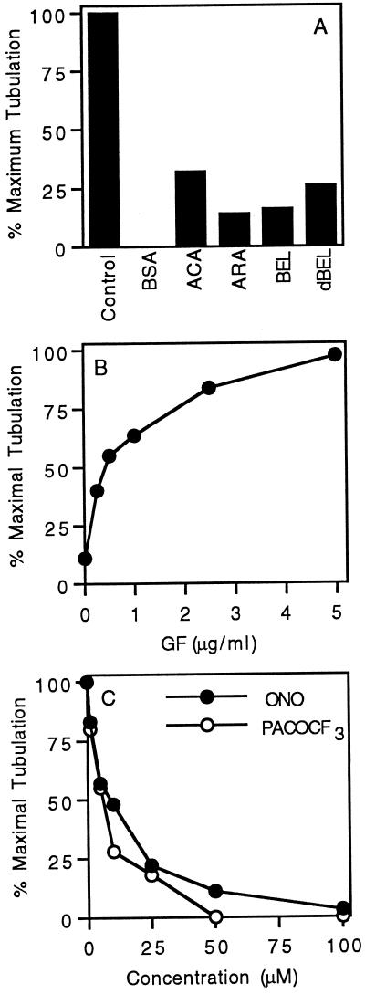
Figure 10.
Quantitation of Golgi membrane tubulation inhibition by PLA2 antagonists. (A) Aliquots of an active fraction of bovine brain cytosol, containing tubulation activity, were incubated in the absence or presence of various PLA2 inhibitors, and then each was mixed with isolated rat liver Golgi complexes in our standard in vitro tubulation assay. Concentrations were BBC (bovine brain cytosol), 1.5 mg/ml; BSA, 1.5 mg/ml; ACA, ARA, and BEL, 5 μM. The bar labeled dBEL (for dialyzed BEL) shows the level of tubulation that resulted when BBC was first incubated with BEL (20 μM for 15 min at 37°C) and then dialyzed to remove excess, unbound BEL. The extent of tubulation was measured as described in MATERIALS AND METHODS, and the data are expressed as the percent maximal tubulation to compare results from different experiments. (B) Addition of the enriched GF fraction restored tubulation activity toBEL-inactivated cytosol. BBC (1.5 mg/ml) was incubated with BEL (as above), dialyzed extensively, incubated with increasing amounts of enriched GF fraction, and then used in the standard in vitro tubulation assay. The data points are averages of duplicate experiments. (C) Typical dose–response experiment showing the loss of cytosolic tubulation activity with increasing amounts of the PLA2 inhibitors ONO-RS-082 and PACOCF3. All data points are the averages of duplicate experiments.

