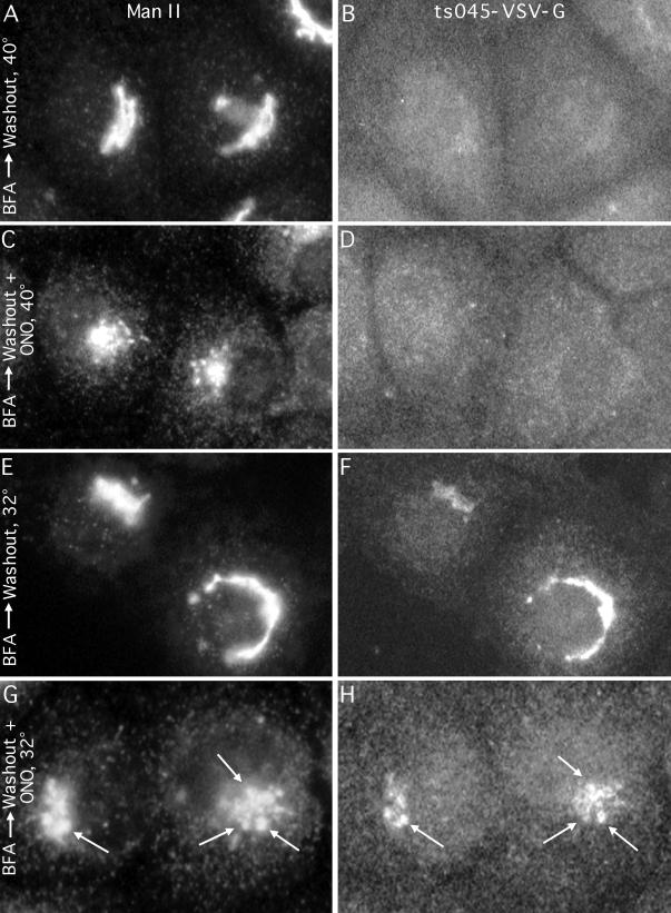
Figure 8.
Golgi mini-stacks formed in the presence of PLA2 inhibitors during reassembly after BFA washout receive membrane traffic from the ER. Cells were infected with ts045 VSV at 40°C to entrap VSV-G in the ER, treated with BFA (5 μg/ml for 15 min) to recycle the Golgi back to the ER, washed free of BFA, and then incubated under various conditions to allow Golgi reassembly and/or transport of VSV-G out of the ER. Cells were then fixed and processed for double-label immunofluorescence to localize ManII (left panels) and VSV-G (right panels). Conditions during the washout from BFA were as follows. (A and B) Cells incubated with drug-free media at 40°C allow the Golgi complex to completely reform, but VSV-G remains diffusely in the ER. (C and D) Cells incubated in media with ONO-RS-082 (ONO) at 40°C reassemble the Golgi into punctate mini-stacks, but VSV-G still remains in the ER. (E and F) Cells incubated with drug-free media but shifted to the permissive temperature of 32°C reassemble an intact Golgi complex to which VSV-G is now transported, as evidenced by its colocalization with ManII. (G and H) Cells incubated with ONO-RS-082 at 32°C reassemble the Golgi only to the point of forming punctate mini-stacks to which VSV-G is transported (arrows point to double-labeled mini-stacks).

