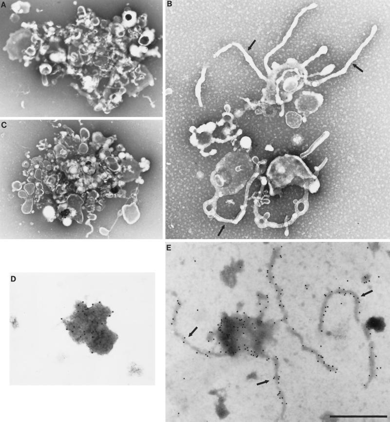
Figure 9.
Electron microscopic observations of Golgi membrane tubulation in a cytosol-dependent, in vitro reconstitution system and immunogold localization of ManII on Golgi membrane tubules. Golgi-enriched fractions were incubated in vitro under various conditions and applied as whole-mount preparations onto EM grids for standard negative staining (A–C) or for a modified protocol involving a combination of immunogold labeling of ManII followed by negative staining (D and E). (A) Golgi complex incubated with buffer control. (B) Golgi complex incubated with bovine brain cytosol under conditions that induce membrane tubule formation. Arrows indicate a few of the numerous 60- to 80-nm-diameter membrane tubules that formed. (C) Golgi complex incubated with cytosol that had first been treated with the PLA2 inhibitor BEL (25 μM). (D) Control Golgi complex immunogold labeled to show distribution of ManII. (E) Golgi complexes incubated with cytosol under tubulation conditions and then immunolabeled to show ManII distribution. Arrows indicate several tubules with a nearly uniform distribution of gold particles along the length of each tubule. As shown in E, all of the induced tubules were labeled with anti-ManII antibodies; however, in many other cases, only about half of the Golgi tubules were labeled with ManII antibodies, and in double-labeling experiments that localized ManII and mannose 6-phosphate receptors (located in trans elements), separate tubules were stained. Bar, 0.5 μm.

