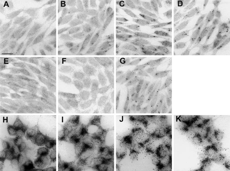
Figure 7.
InsP3 receptor clustering in E36M3R and AR4–2J cells. Cells were stimulated, fixed, and permeabilized and then incubated with antibodies against type II or III InsP3 receptors and rhodamine-conjugated secondary antibodies. (A–G) E36M3R cells stained for type III receptor after exposure to 1 mM carbachol for 0 min (A), 10 min (B), 30 min (C), and 60 min (D) or for 60 min to 1 mM carbachol plus 10 μM atropine (E). Alternatively, E36M3R cells were treated with 400 nM PMA (F) or 2 μM thapsigargin (G). (H–K) AR4–2J cells stained for type II receptor after exposure to 0.5 μM CCK for 0 min (H), 10 min (I), 30 min (J), and 60 min (K). Micrographs shown are representative of at least two independent experiments. Bar, 20 μm.

