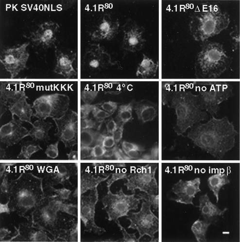
Figure 5.
In vitro nuclear import assay of PK and 4.1R80 in permeabilized COS-7 cells. Subconfluent COS-7 cells grown on coverslips were permeabilized in import buffer containing 50 μg/ml digitonin and washed twice in import buffer. Cells were then incubated with recombinant import substrates together with GTP, an ATP regeneration system, and recombinant importin α2 (Rch1), importin β, and GTPase Ran. Import substrates include PK/SV40NLS, 4.1R80, 4.1R80ΔE16, and 4.1R80mutKKK (first four panels). Control experiments showed that import was blocked by incubation at 4°C, by preincubation with WGA, by omission of GTP and the ATP regeneration system, or by omission of Rch1 or importin β (remaining panels). Cells were fixed in 3% paraformaldehyde and permeabilized in 0.5% Triton X-100. Samples were then processed for immunofluorescence using an affinity-purified polyclonal antibody against the S tag as primary antibody and anti-rabbit IgG coupled to FITC as secondary antibody. Specificity of detection was assessed using samples in which primary S-tag antibody was either omitted or pre-exhausted with control peptide. Cell imaging was performed on samples analyzed by conventional microscopy. The pattern of cells probed with wild-type PK was similar to that of 4.1R80 mutants. Bar, 10 μm.

