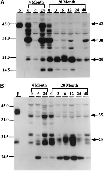
Figure 4.
Western blot analysis of the levels of C/EBPα and C/EBPβ isoforms in liver nuclei in response to LPS treatment. Nuclear extracts (30 μg) from control (0) and LPS-injected (3, 6, 12, 24, and 48 h postinjection) C57BL/6 mice (4- and 28-mo-old males) were loaded in each lane and subjected to ECL-Western immunoblot analyses as described in MATERIALS AND METHODS. Immunoblots were incubated with monospecific polyclonal antibodies against C/EBPα (A) or C/EBPβ (B) or preimmune serum as a control (unpublished data). (A) Anti-C/EBPα. (B) Anti-C/EBPβ. Lanes α and β represent nuclear extracts from COS-1 cells transfected with C/EBPα or C/EBPβ expression vectors and were used as standards for each protein. No bands were detected with preimmune serum with liver nuclear extracts or COS-1 nuclear proteins (unpublished data). Positions of molecular mass standards (kDa) are indicated on the left.

