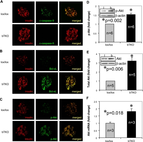
Figure 8.
Induction of Akt/Bcl-xL signaling in bTKO mice. Control lox/lox and bTKO mice received multiple low-dose STZ injections; 8 days after the initial injection, pancreata were analyzed by immunohistochemistry for cleaved caspase-9 (A), Bcl-xL (B), and p-Akt (C). Using isolated islets of untreated lox/lox and bTKO mice, the induction of Akt was further quantified by immunoblotting for p-Akt (D) and total Akt (E) and by quantitative real-time RT-PCR for Akt mRNA (F). Bars represent means ± se; n = mice/group. Insert: representative immunoblot (D, E).

