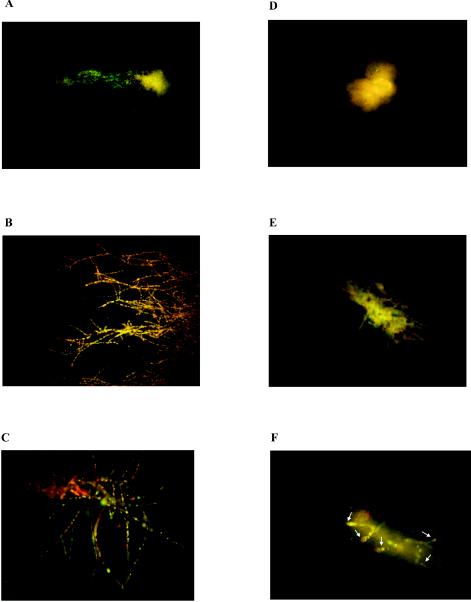
FIG. 1.

FUN-1 staining of A. fumigatus hyphae. Panels A to C show damage induced by ITC at a concentration of 2 μg/ml to hyphae in A. fumigatus isolate 32 after four passages on YAG plates containing no FLC (A), 8 μg of FLC/ml (B), or 256 μg of FLC/ml (C). Panels D to F show damage induced by ITC at a concentration of 256 μg/ml to hyphae in A. fumigatus isolate 32 after four passages on YAG plates containing no FLC (D), 8 μg of FLC/ml (E), or 256 μg of FLC/ml (F). Arrows (F) indicate viable hyphae. Compartments of Aspergillus hyphae previously exposed to FLC remained viable even after exposure to ITC at a concentration of 256 μg/ml.
