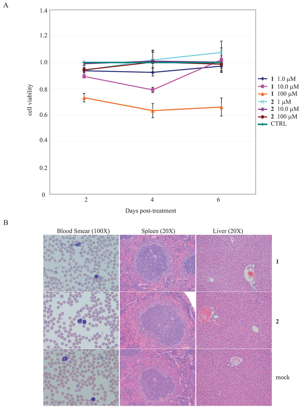
Figure 6.
Lack of toxicity of 1 and 2 in cells and in mice. A) 4 × 10⁶ MT4 cells in 2 ml were incubated in culture media containing 1, 10, and 100 µM concentrations of 1 and 2. 100 µl of each cell suspension were incubated with 10 µl of WST8 (water-soluble tetrazolium) for 2 hrs and then subjected to colorimetric measurements. Production of color is a measurement of the metabolism of viably dividing cells. B) In vivo toxicities of 1 and 2 were evaluated in 8–10 week old Balb-C mice. Mice, in groups of 4, were injected intraperitoneally twice weekly with drug boluses calculated to achieve final body 1 and 2 distributions of 0.2, 2, 20 and 200 µM, respectively for 4 weeks. None of the mice demonstrated any signs of toxicity from intra-peritoneal injections of 1 or 2. At the end of 5 weeks of drug injection and observation, all mice were sacrificed and histological examinations of tissues were performed. Examples of tissues from 1 and 2 injected mice compared to tissues from control mice are shown.

