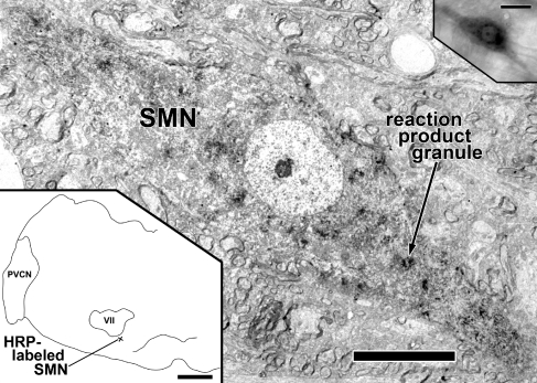
FIG. 1.
HRP-labeled SMN. Center image: low magnification electron micrograph of SMN (neuron R1) with reaction product granules (one indicated) and diffuse darkening of the cytoplasm. Lower left: drawing of the left half of a coronal section of a rat brainstem showing the location of the SMN ventromedial to the left motor nucleus of the facial nerve (VII). The pyramidal tract is outlined at the midline. PVCN posteroventral cochlear nucleus. Upper right: bright-field photomicrograph of the epoxy-embedded labeled SMN in an 80-μm thick Vibratome section. Scale bar = 10 μm (center image), scale bar = 1 mm (lower left), scale bar = 25 μm (upper right).

