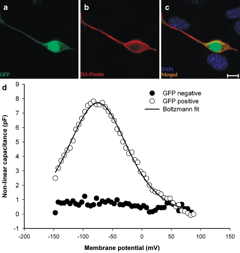
FIG. 2.
GFP and HA–prestin expression in HEK 293 cells. A Transduced cells were identified by GFP expression. B HA–prestin immunolabeling was detected in the HEK cell membrane. C A merged image with DAPI nuclear staining demonstrated that only transduced cells expressed GFP and HA–prestin. D Example capacitance versus voltage curves, after subtracting the linear capacitance, in both a GFP-positive cell and a control cell in which HDAd–prestin–GFP was not applied. A nonlinear capacitance was only identified in GFP-positive cells. The solid black line represented Boltzmann fits to the data. Fit parameters were Vpkcm = −74.8 mV, α = 33.8 mV, Qmax = 1040.9 fC. (A), (B), and (C) bar = 10 μm.

