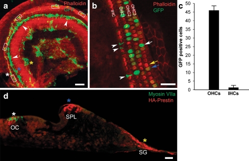
FIG. 3.
GFP and HA–prestin expression within cochlear sensory epithelium cultures from prestin-null mice. A A confocal image of a whole-mount preparation under low magnification demonstrates that GFP was expressed primarily within the organ of Corti area (white asterisk and rectangles), the spiral limbus (blue asterisk), and the spiral ganglion area (yellow asterisk). B A representative confocal image under high magnification demonstrates transduction of OHCs and supporting cells. Some OHCs had bright GFP fluorescence (white arrow), some OHCs had low GFP fluorescence (blue arrow), and some OHCs had no GFP fluorescence (yellow arrow). Supporting cells were also transduced (arrowheads). C The percentage of GFP-positive and GFP-negative hair cells was determined in 35 images from 7 cochleae [representative areas counted are shown in (A), rectangles]. D A paraffin-embedded cross-section of a cultured preparation demonstrates that HA–prestin was expressed within the organ of Corti (OC, white asterisk), the spiral limbus (SPL, blue asterisk), and the spiral ganglion area (SG, yellow asterisk). These asterisks correspond to those in (A). Myosin VIIa antibody labels the hair cells with green fluorescence. Scale bars: A 200 μm, B 50 μm, D 100 μm.

