FIG. 4.
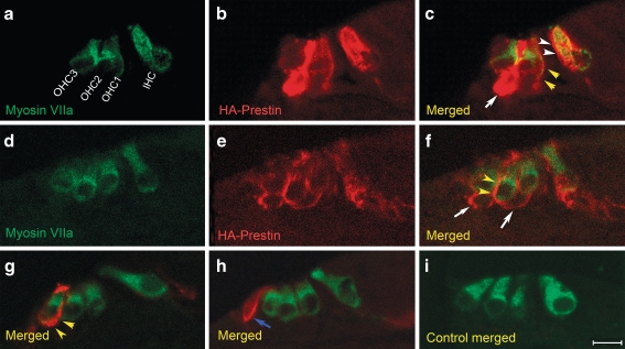
HA–prestin immunolocalization within the organ of Corti of prestin-null cochleae. Selected paraffin-embedded cross-sections of the hair cell region. The orientation is the same for all images, with an IHC on the right and three rows of OHCs on the left. One selected section demonstrates myosin VIIa immunolabeling (green) to identify hair cells (A), HA immunolabeling (red) to identify prestin (B), and a merged image (C). In this example, HA–prestin was expressed in IHCs (white arrowheads), OHCs (yellow arrowheads), and Deiter cells (white arrows). The HA–prestin labeling was primarily within the plasma membrane. D, E, F Another selected section demonstrates HA–prestin expression in OHCs (yellow arrowheads) and Deiter cells (white arrows). Two more selected sections are presented with myosin VIIa and HA–prestin immunolabeling fluorescence merged. A single OHC (G, yellow arrowheads), and a single Hensen cell (H, blue arrow) demonstrated HA–prestin expression. I A control organ of Corti demonstrated no HA–prestin immunolabeling. Scale bar: A–I 10 μm.

