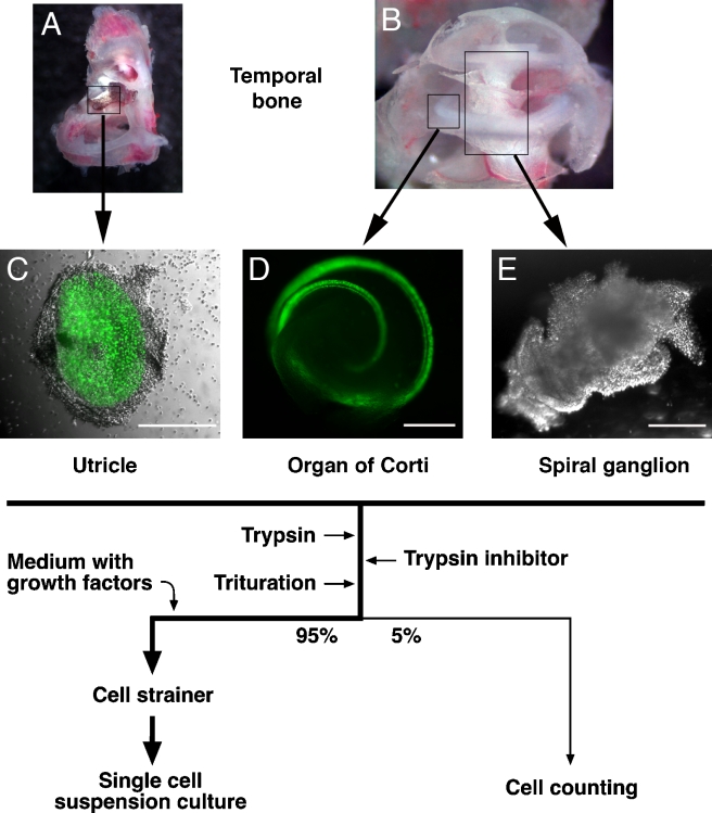
FIG. 1.
Tissue isolation and cell handling procedure. A Temporal bone with utricle (box). B Cochlea. Positions of a sector of the organ of Corti (small box) and the spiral ganglion (large box) are indicated. C–E Dissected utricular macula, organ of Corti, and spiral ganglion. nGFP-positive hair cells are visualized by green fluorescence. Flow chart, generation of single cell suspension cultures. Images show specimen dissected at 0 h postmortem from 3-week-old (A, B) and newborn (C–E) mice. Scale bars = 300 μm.

