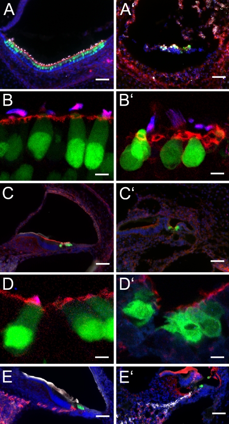
FIG. 2.
Cross-sections through the inner ear of newborn Math1/nGFP mice at 0 h (A–E) and 10 days postmortem (A′–E′). A, A′ Shown is the utricle with nGFP-positive hair cells (green). Filamentous actin is visualized in red, and the hair bundle protein espin, detected with Cy5-conjugated secondary antibody, is depicted in white. Nuclei are visualized with DAPI (blue). B, B′ Higher magnification of utricular hair cells reveals loss of nuclear confinement of nGFP (green) at 10 days postmortem. Hair bundles are visualized with antibody to espin (blue) and by labeling with rhodamine-conjugated phalloidin (red) to detect filamentous actin. C, C′ Organ of Corti sections and D, D′ higher magnification images of cochlear hair cells. E, E′ In the spiral ganglion, we observed massive loss of β-III tubulin (TuJ)-immunopositive neural cell bodies and their associated neurites (depicted in red). GFAP immunoreactivity (shown in white) was present at 0 h, although not very pronounced, but appeared stronger at 10 days postmortem than at 0 h. Note that the labeling pattern of antibodies to TuJ and to GFAP appears to be altered at 10 days postmortem, possibly due to extensive autolytic processes that probably reveal novel unspecific antigens in the tissue. The apparent positive immunoreactivity of the tectorial membrane is unspecific because it is also detectable in negative controls where the primary antibodies were omitted. nGFP-positive hair cells are shown in green and nuclei are visualized with DAPI (shown in blue). Scale bars = 50 μm in A, A′, C, C′, E, and E′ and 10 μm in B, B′, D, and D′.

