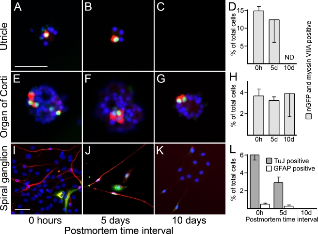
FIG. 7.
Differentiation of mature cell types from spheres derived at different postmortem time intervals. (A–B) Hair-cell-like cells, defined by nGFP expression (green) and labeling with antibody to myosin VIIA (red) can be found in differentiated cells from utricle-derived spheres at 0 h and 5 days postmortem. C At 10 days postmortem, utricle-derived spheres failed to attach to the substrate. D Quantitative assessment of the number of nGFP and myosin VIIA double positive cells of the total cell number of differentiated utricle-derived spheres. E–G Occurrence of hair-cell-like cells in organ of Corti-derived spheres at 0 h and 5 and 10 days postmortem. H Quantitative assessment of the number of nGFP and myosin VIIA double positive cells of the total cell number of differentiated organ of Corti-derived spheres. I–K Neurons, defined by expression of β-III tubulin (TuJ, shown in red), and GFAP expressing cells (shown in green) can be found in differentiated cells from spiral ganglion-derived spheres at 0 h and 5 days postmortem, but not at 10 days postmortem. In some instances, we detected very weak expression of TuJ in GFAP-positive cells, which we interpret as a sign of immaturity of these newly generated cells. L Quantitative assessment of the number of TuJ- and GFAP-positive cells of the total cell number of differentiated spiral ganglion-derived spheres. Nuclei are visualized with DAPI (shown in blue); scale bars = 50 μm.

